
Animal Cell Diagram
This animal cell diagram shows the twelve structures that biology courses ask about most, from the cell membrane and nucleus to mitochondria, the Golgi apparatus and both kinds of endoplasmic reticulum. Download it labeled or blank, print it for revision, or copy it into the editor and change any label, line or color.
Labeled Animal Cell Diagram
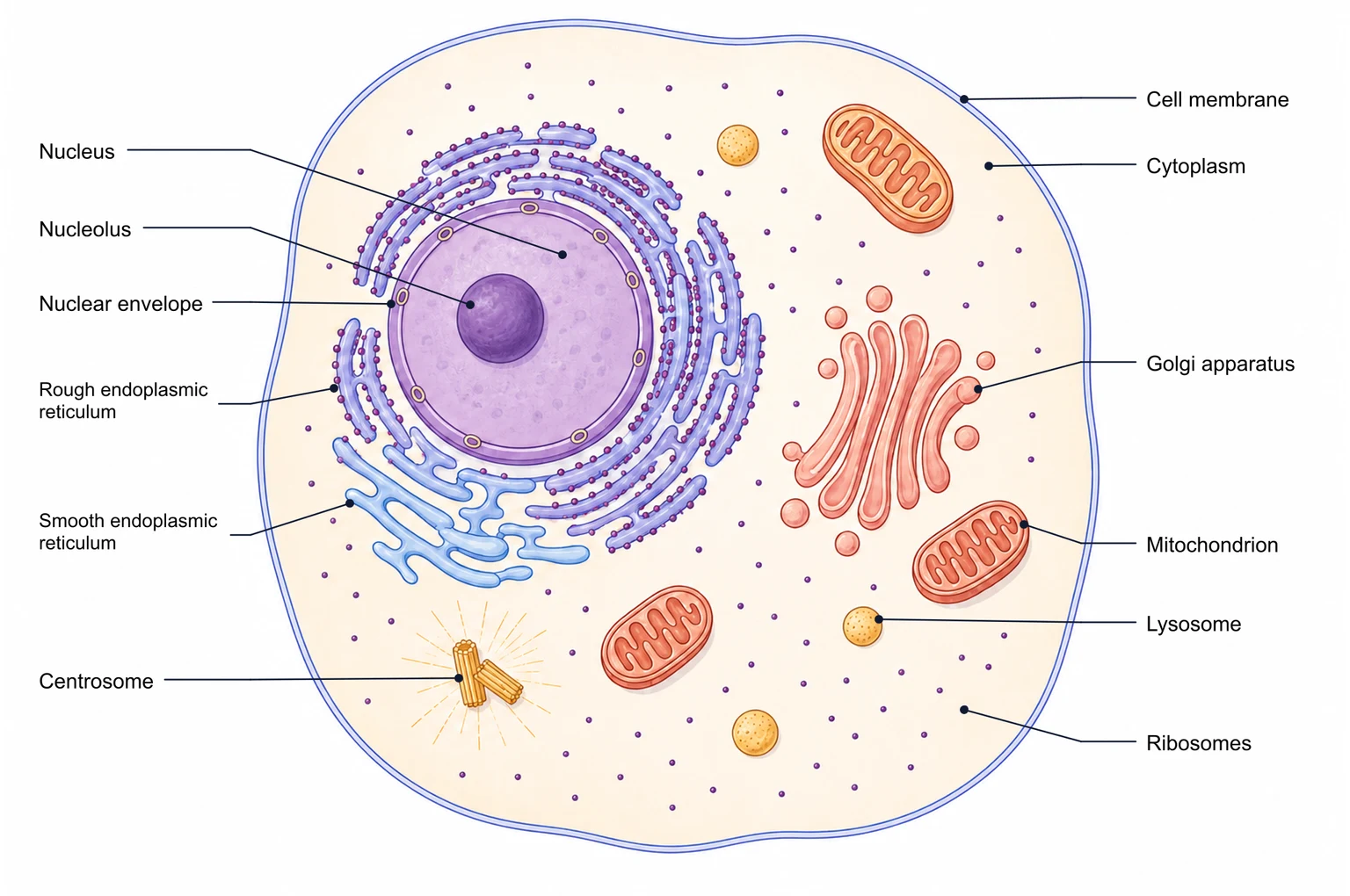
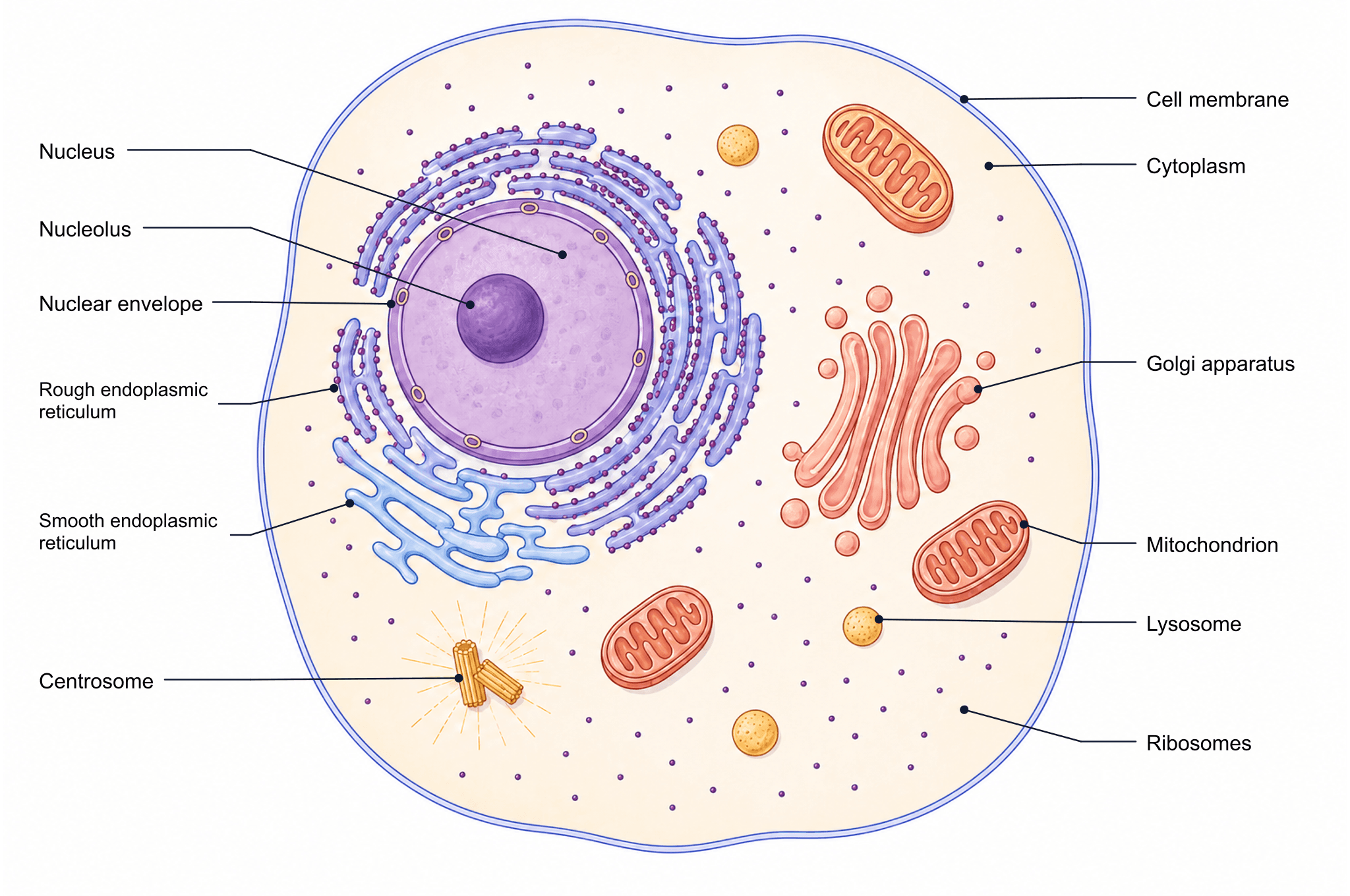
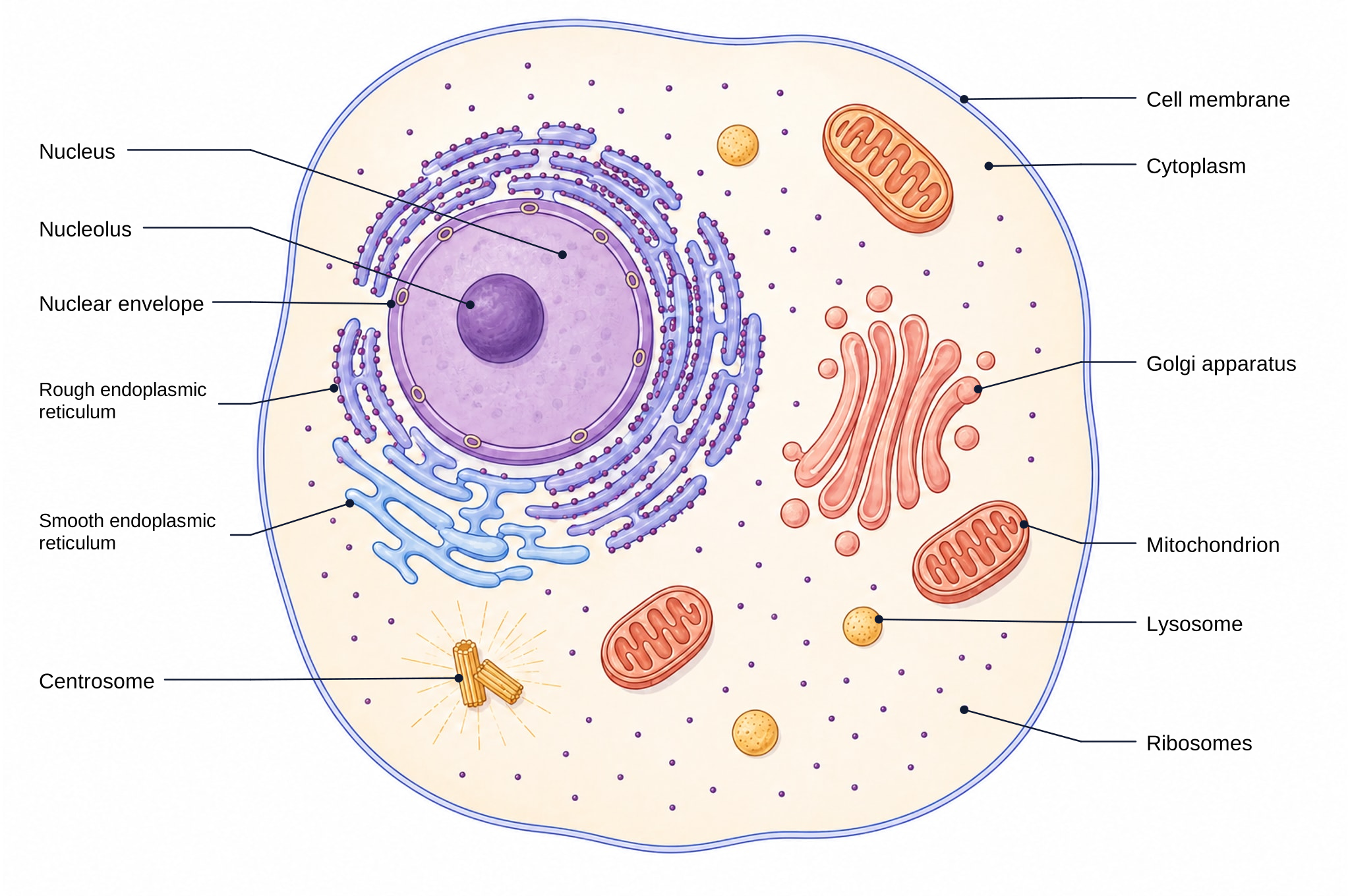
The picture above is an animal cell diagram labeled with twelve structures, drawn as a cross-section so that organelles inside the cell are visible. Shapes follow the conventions of school and first-year university textbooks: a round nucleus with a dark nucleolus, rough ER studded with ribosomes, a stack of Golgi cisternae and bean-shaped mitochondria with folded inner membranes. The table explains what each labeled part does.
| Structure | What it does |
|---|---|
| Cell membrane | A phospholipid bilayer with embedded proteins that encloses the cell and controls which substances enter and leave. |
| Cytoplasm | The gel-like fluid (cytosol) and everything suspended in it outside the nucleus; many metabolic reactions, including glycolysis, take place here. |
| Nucleus | Holds the chromosomes (DNA) and controls the cell by deciding which genes are transcribed into RNA. |
| Nuclear envelope | A double membrane around the nucleus; its nuclear pores let RNA and proteins pass between the nucleus and the cytoplasm. |
| Nucleolus | A dense region inside the nucleus where ribosomal RNA is made and ribosome subunits are assembled. |
| Rough endoplasmic reticulum | Flattened membrane sacs covered with ribosomes; it makes and folds proteins that will be secreted or placed in membranes. |
| Smooth endoplasmic reticulum | Tubular membranes without ribosomes that make lipids and steroids, store calcium ions and help detoxify drugs. |
| Golgi apparatus | A stack of membrane sacs that modifies, sorts and packages proteins and lipids into vesicles for transport or secretion. |
| Mitochondrion | Site of aerobic respiration; the folded inner membrane (cristae) produces most of the cell’s ATP. |
| Lysosome | A small vesicle of digestive enzymes that breaks down worn-out organelles, food particles and engulfed bacteria. |
| Ribosomes | Tiny particles of RNA and protein that translate messenger RNA into protein; found free in the cytoplasm and on the rough ER. |
| Centrosome | Contains a pair of centrioles at right angles and organizes the microtubules that form the spindle during cell division. |
Animal Cell Diagram and Labels for Each Organelle
Labels sit outside the cell with thin leader lines, so nothing covers the structure it names. If you need fewer labels for a younger class, or extra ones such as vacuoles or the cytoskeleton, edit the copy in your account.
Blank Animal Cell Diagram for Practice
Switch to the Blank tab for the same drawing with the label text removed. Print it, cover the table, and test yourself by writing each name at the end of its line.
Plant and Animal Cell Diagram Compared
A diagram of the animal cell and plant cell side by side shows the differences: plant cells add a cell wall, chloroplasts and one large central vacuole, while animal cells have centrioles, lysosomes and no wall, so their shape is more irregular.
Why Use Our Animal Cell Diagram?
Most pictures you find online are fixed images: the labels cannot be changed, the colors may not suit your slides, and the resolution is often too low to print. Students often search for a cell diagram of animal and plant cells that they can adapt; this one is built to be reused.
Edit Labels and Colors
Copy the diagram into your account and the labels become real text: rename a structure, translate every label into another language, change font size, or recolor organelles to match a worksheet or lecture theme. You can also describe a new figure and generate it from scratch.
Create your own figurePrint or Use in Slides
The PNG is 2048 pixels wide, sharp enough for an A4 handout or a full-screen slide. The SVG keeps the labels as text, so it stays crisp when you scale it in PowerPoint, Keynote or Google Slides.
How to Customize the Diagram in 3 Steps
Downloading needs no account. To change the drawing itself, sign in once and the editor opens with your own copy.
Copy it to your account
Click "Customize this diagram". A copy of the labeled version, with every label as editable text, opens in the editor.
Change labels, lines and colors
Double-click a label to rewrite it, drag leader lines to new positions, delete what you do not need, or apply a color palette to the organelles.
Export for class or a paper
Export PNG for documents, SVG for further editing, or PPTX with labels as separate objects for teaching slides.
Animal Cell Diagram FAQ
Related Tools