
Neuron Diagram
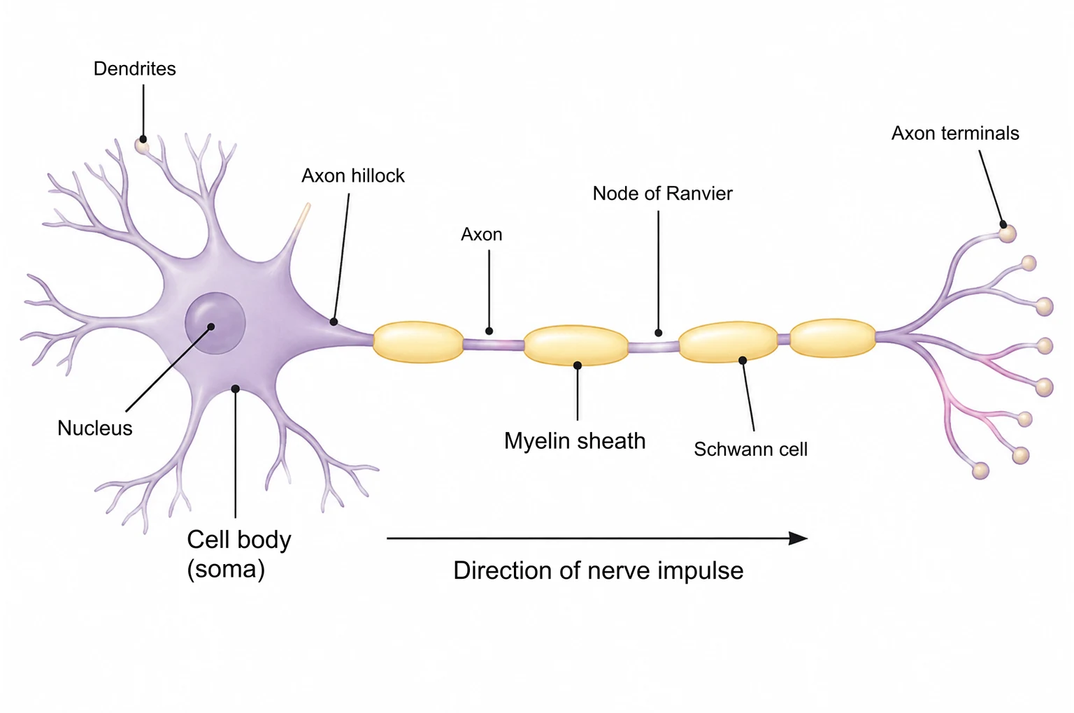
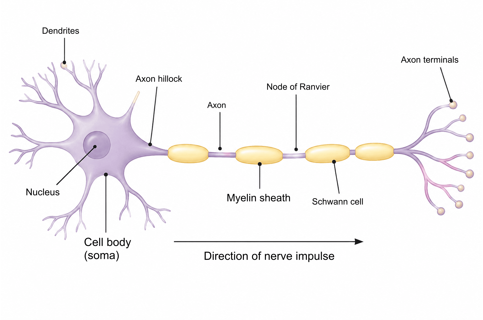
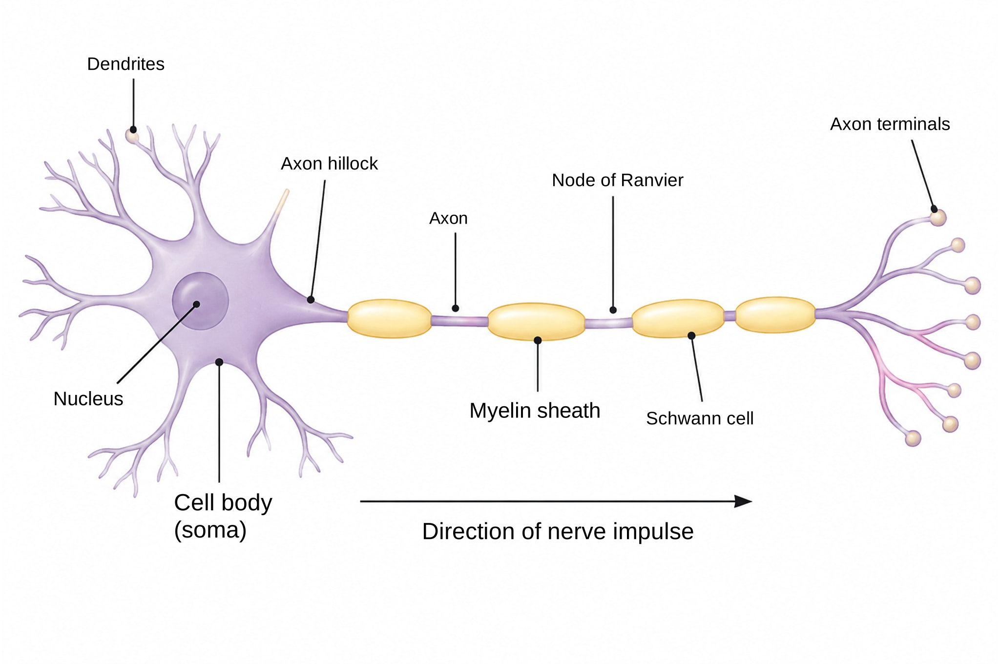
This neuron diagram shows a myelinated nerve cell from end to end: branching dendrites, the cell body with its nucleus, the axon hillock, an axon wrapped in myelin with nodes of Ranvier, and the axon terminals, with an arrow for the direction of the nerve impulse. Download it labeled or blank, or open your own copy in the editor and change any label.
Labeled Neuron Diagram
The drawing above is a neuron diagram labeled with ten items, laid out left to right in the order a signal travels: it is received by the dendrites, summed at the cell body, started at the axon hillock, conducted along the axon and passed on at the terminals. The table gives each part a one-line function you can learn from.
| Part | Function |
|---|---|
| Dendrites | Short, branched extensions that receive chemical signals from other neurons and carry them toward the cell body. |
| Cell body (soma) | Contains the nucleus and most organelles; it keeps the cell alive and combines the incoming signals. |
| Nucleus | Holds the DNA and directs the production of proteins, including neurotransmitter-making enzymes and ion channels. |
| Axon hillock | The cone-shaped region where the axon leaves the soma; if the summed input reaches threshold, the action potential starts here. |
| Axon | A long, thin fibre that conducts the action potential away from the cell body, sometimes for more than a metre. |
| Myelin sheath | Layers of insulating membrane around the axon that speed up conduction and reduce the energy each impulse costs. |
| Schwann cell | The glial cell that wraps around the axon to form each myelin segment in the peripheral nervous system (oligodendrocytes do this in the brain and spinal cord). |
| Node of Ranvier | A short gap between myelin segments, rich in sodium channels, where the impulse is regenerated so that it jumps from node to node (saltatory conduction). |
| Axon terminals | Branched endings that release neurotransmitter from vesicles into the synapse with the next neuron, muscle or gland. |
| Direction of nerve impulse | The arrow shows one-way travel: from dendrites through the soma and along the axon to the terminals. |
Neuron Diagram Labelled Parts and Functions
British and Australian courses spell it "labelled"; the parts are the same. Every label sits beside the structure with a thin leader line, so the drawing stays clear when you print it in black and white.
Motor Neuron Diagram
The shape drawn here is the classic motor neuron diagram: many dendrites around a large cell body, and one long myelinated axon ending in terminals that would meet muscle fibres. Sensory and relay neurons put the cell body in other positions. In the spinal cord, the cell body of a motor neuron sits in the ventral horn, and its axon leaves through the ventral root to reach a skeletal muscle.
Neuron Synapse Diagram
This drawing stops at the axon terminals. At a synapse, each terminal releases neurotransmitter across a narrow gap, the synaptic cleft, onto receptors in the postsynaptic membrane of the next cell. Describe that scene in the generator if you need a separate synapse figure.
Why Use Our Neuron Diagram?
A diagram of a neuron is reused again and again: in revision notes, lab reports, lecture slides and exam practice. Each use needs something slightly different, such as fewer labels, another language or a color that matches your slides, so this one is built to be changed.
Edit Labels for Your Course
In your own copy, every label is real text. Rename "cell body (soma)" to match your textbook, add "oligodendrocyte" for a central nervous system version, or delete labels to make a quiz. You can also generate a new figure from a description.
Create your own figureUse It in Slides or Print
The PNG is 2048 pixels wide, enough for a full-screen slide or an A4 handout. The SVG keeps labels as text for clean scaling. We made a matching labeled animal cell drawing in the same style.
See the animal cell diagramHow to Customize the Diagram in 3 Steps
Downloads need no account. To edit the drawing, sign in once and your copy opens in the editor.
Copy the diagram
Click "Customize this diagram" to put a copy of the labeled version in your account and open it.
Edit labels, lines and colors
Rewrite a label by double-clicking it, move or delete leader lines, or change colors with a palette.
Export it
Download PNG for documents, SVG for scaling, or PPTX with each label as its own object for slides.
Neuron Diagram FAQ
Related Tools